|
| |

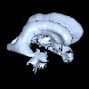
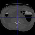
Head MRT Angiography 16Bits
 Head Angiography Dataset provided by Volvis Project Group of Tuebingen University, Germany.
3T MRT Time-of-Flight Angiography dataset of a human head. The dataset has been resampled into an isotropic voxel grid (hence the peculiar slice size). The 8 bits version can be found here.
File Format: raw
Bits per Voxel: 16Bits (10bits set)
Size:416 x 512 x 112
Spacing (mm): 0.412, 0.412, 0.412
Courtesy of: Özlem Gürvit, Institute for Neuroradiology, Frankfurt, Germany.
Original Page: University of Tübingen Datasets Page
|
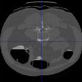
Head MRT Angiography 16Bits Segmented
Head Angiography Dataset provided by Volvis Project Group of Tübingen University, Germany.
3T MRT Time-of-Flight Angiography dataset of a human head. The dataset has been resampled into an isotropic voxel grid.
Also a segmentation of the arterial blood vessels is provided here, where the first (negative) number is the number of all voxels of the segmentation, followed by the voxel array indices of the voxels of the segmentations.
The 8 bits version can be found here.
File Format: raw
Bits per Voxel: 8Bits
Size: 256 x 320 x 128
Spacing (mm): 0.66, 0.66, 0.66
Courtesy of: Özlem Gürvit, Institute for Neuroradiology, Frankfurt, Germany. For the segmentation: Dirk Bartz, VCM, University of Tübingen, Germany.
Original Page: University of Tübingen Datasets Page
|
Head MRI CISS 8Bits
1.5T MRT 3D CISS dataset of a human head that highlights the CSF (Cerebro-Spinal-Fluid) filled cavities of the head. Also a segmentation of the cerebral ventricular system is provided here, where the first (negative) number is the number of all voxels of the segmentation, followed by the voxel array indices of the voxels of the segmentations.
File Format : raw
Bits per Voxel : 8 Bits
Size : 256 x 256 x 124
Spacing (mm) : 0.9, 0.9, 0.9
Courtesy of: Dirk Bartz, VCM, University of Tübingen, Germany.
Original Page: University of Tübingen Datasets Page
|
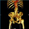
Stented Abdominal Aorta 16Bits
CT Scan of the abdomen and pelvis. The dataset contains also a stent in the abdominal aorta. No contrast agent was used to enhance the blood vessels.
File Format : raw
Bits per Voxel : 16 Bits
Size : 512 x 512 x 174
Spacing (mm) : 0.8398, 0.8398, 3.2
Courtesy of: Michael Meißner, Viatronix Inc., USA.
Original Page: University of Tübingen Datasets Page
|
Head Aneuyrism 16Bits
Vertebra Dataset provided by Volvis Project Group of Tübingen University, Germany.
Rotational angiography scan of a head with an aneurysm. Only contrasted blood vessels are visible.
The 8 bits version can be found here.
File Format : raw
Bits per Voxel : 16 Bits
Size : 512 x 512 x 512
Spacing (mm) : 0.1953, 0.1953, 0.1953
Courtesy of: Michael Meißner, Viatronix Inc., USA.
Original Page: University of Tübingen Datasets Page
|
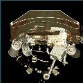
Backpack Scan 16Bits
CT scan of a backpack filled with items. The 8 bits version can be found here.
File Format: raw
Bits per Voxel: 16 Bits (12 bits set)
Size: 512 x 512 x 373
Spacing (mm): 0.9766, 0.9766, 1.25
Courtesy of: Kevin Kreeger, Viatronix Inc., USA..
Original Page: University of Tübingen Datasets Page
|
Colon Prone 16 Bits
CT scan of abdomen in prone orientation, back faces ceiling, belly faces table.
The 8bits version can be found here.
File Format: raw
Bits per Voxel: 16 Bits (12 bits set)
Size: 512 x 512 x 463
Spacing (mm): 0.625, 0.625, 1.0
Courtesy of: Walter Reed Army Medical Center, USA.
Original Page: University of Tübingen Datasets Page
|
Colon Supine 16 Bits
CT scan of abdomen in supine orientation (back faces table, belly faces ceiling. The 8 bits version can be found here.
File Format : raw
Bits per Voxel : 16Bits (12bits set)
Size : 512 x 512 x 426
Spacing (mm) : 0.625, 0.625, 1.0
Courtesy of: Dirk Bartz, VCM, University of Tübingen, Germany.
Original Page: University of Tübingen Datasets Page
|
Colon Phantom 16 Bits
CT scan of a Colon phantom with several different objects and five pedunculated large polyps in the central object. The 8 bits version can be found here.
File Format : raw
Bits per Voxel : 16Bits (12bits set)
Size : 512 x 512 x 442
Spacing (mm) : 0.9316, 0.9316, 0.5
Courtesy of: Michael Meißner, Viatronix Inc., USA..
Original Page: University of Tübingen Datasets Page
|

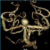
Aneurism
Rotational C-arm x-ray scan of the arteries of the right half of a human head. A contrast agent was injected into the blood and an aneurism is present..
File Format : raw
Bits per Voxel : 8Bits
Size : 256x256x256
Spacing (mm) : 1, 1, 1
Courtesy of: Philips Research, Hamburg, Germany
Original Page: University of Tübingen Datasets Page
|
|