2.2. THE HUMAN VISUAL
SYSTEM
2.2.1. The Eye
The human visual system is one of the most complex in existence. Our visual system allows us to
organize and understand the many complex elements of our environment. For
nearly all animals, vision is just an instrument of survival. For humans,
vision is not only an aid to survival, but an instrument of thought and a means
to a richer life.
The visual system consists of an eye that
transforms light to neural signals, and the related parts of the brain that
process the neural signals and extract necessary information. The eye, the
beginning of the visual system, is approximately spherical with a diameter of
around 2 cm. From a functional point of view, the eye is a device that gathers
light and focuses it on its rear surface.
A horizontal cross section of an eye is
shown in Figure 2.10. At the very front of the eye, facing the outside world, is the cornea, a tough, transparent membrane. The
main function of the cornea is to refract (bend) light. Because of its rounded
shape, it acts like the convex lens of a camera. It accounts for nearly
two-thirds of the total amount of light bending needed for proper focusing.
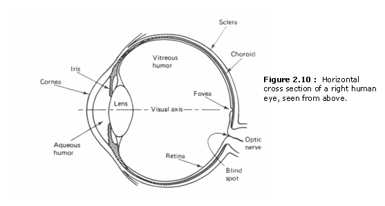
Behind the cornea is the aqueous humor, which is a clear, freely flowing
liquid. Through the cornea and the aqueous humor, we can see the iris. By
changing the size of the pupil, a small round hole in the center of the iris,
the iris controls the amount of light entering the eye. Pupil diameter ranges
between 1.5 mm ~ 8 mm, with smaller diameter corresponding to exposure to
brighter light. The color of the iris determines the color of the eye. When we
say that a person has blue eyes, we mean blue irises. Iris color, which has
caught the attention of so many lovers and poets, is not functionally
significant to the eye.
Behind the iris is the lens. The lens consists of many transparent
fibers encased in a transparent elastic membrane about the size and shape of a
small bean. The lens grows throughout a person’s lifetime. Thus, the lens of an
eighty-year-old man is more than fifty percent larger than that of a
twenty-year old. As with an onion, cells in the oldest layer remain in the
center, and cells in newer layers grow further from the center. The lens has a
hi-convex shape and a refractive index of 1.4, which is higher than any other
part of the eye through which light passes. However, the lens is surrounded by
media that have refractive indices close to its own. For this reason, much less
light-bending takes place at the lens than at the cornea. The cornea has a
refractive index of 1.38, but faces the air, which has a refractive index of 1.
The main function of the lens is to accurately focus the incoming light on a
screen at the back of the eye called the retina. For a system with a fixed lens
and a fixed distance between the lens and the screen, it is possible to focus
objects at only one particular distance. if faraway
objects are in sharp focus, for example, close objects will be focused behind
the screen. To be able to focus close objects at one time and distant objects
at some other time, a camera changes the distance between the fixed lens and
the screen. This is what the eyes of many fish do. in
the case of the human eye, the shape of the lens, rather than the distance
between the lens and screen, is changed. This process of changing shape to meet
the needs of both near and far vision is called accommodation. This adjustability of the shape is the most
important feature of the lens. Accommodation takes place almost instantly and
is controlled by the ciliarv body, a group of muscles
surrounding the lens.
Behind the lens is the vitreous humor, which is a transparent jelly-like
substance. It is optically matched so that light which has been sharply
focused by the lens keeps the same course. The vitreous humor fills the entire
space between the lens and the retina and occupies about two-thirds of the
eye’s volume. One of its functions is to support the shape of the eye.
Behind the vitreous humor is the retina, which covers about 65% of the
inside of the eyeball. This is the screen on which the entering light is
focused and light-receptive cells convert light to netural
signals. All of the other eye parts we have discussed so far serve the function
of placing a sharp image on this receptor surface. The fact that an image is
formed on the retina, so the eye is simply an image catching device, was not
known until the early seventeenth century. Even though the ancient Greeks knew
the structure of an eye accurately and performed delicate surgery on it, they
theorized that light-like rays emanate from the eye, touch an object, and make
it visible. After all, things appeal “out there” In 1625. Schemer demonstrated
that light enters the eye and vision stems from the light that enters the eye.
By exposing the retina of an animal and looking through it from behind. he was able to see miniature reproductions of the objects in
front of the eyeball.
There are two types of light-receptive cells in the retina. They are
called cones and rods because of their shape. The cones, which number about 7
million, are less sensitive to light than rods, and are primarily for day (photopic) vision. They are also responsible for seeing
color. The three types of cones are most sensitive to red, green, and blue
light, respectively. This is the qualitative phvsiological
basis for representing a color image with red, green. and
blue monochrome images. The rods, which number about 120 million, are more
sensitive to light than cones, and are primarily for night (scotopic)
vision. Since the cones responsible for color vision do not respond to dim
light, we do not see color in very dim light.
Rods and cones are distributed throughout the retina. However, their distribution
is highly uneven. The distribution of the rods and cones in the retina is shown
in Figure 2.11. Directly behind the middle point of the pupil is a small
depressed dimple on the retina called the fovea. There
are no rods in this small region, and most of the cones are concentrated here.
Therefore, this is the regiot for the most accurate
vision in bright light. When we look straight ahead at a,’ object, the object
is focused on the fovea. Since the fovea is very small, we constantly move our
attention from one region to another when studying a larger region in detail.
The rods, which function best in night vision, are concentrated away from the
fovea. Since there are no rods in the fovea, an object focused in the fovea is
not visible in dim light. To see objects at night, therefore, we look at them
slightly sideways.
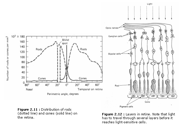
There are many thin layers in the retina. Even though cones and rods arc
light-receptive cells, so that it would be reasonable for them to be located closei to the vitreous humor, they are located farther away
from it. Therefore, light has to pass through other layers of the retina, such
as nerve fibers, to reach the cones and rods. This is shown in Figure 2.12. it is not clear why nature chose to do it this way, but the
arrangement works. in the fovea, at least, the nerves
are pushed aside so that the cones are directly exposed to light. Due to this
particular arrangement, the optic nerve fibers have to pass through the
light-receptive cell layers
on the way to the brain, instead of
crossing the light-receptive cell layers throughout the retina, they bundle up
at one small region the size of a pinhead in the retina, known as the blind
spot. Since there are no light receptive cells in this region, we cannot see
light focused on the blind spot.
When light hits cones and rods, a complex electrochemical reaction takes
place, and light is converted to neural impulses, which are transmitted to the
brain through the optic nerve fibers. There are about 130 million
light-receptive cells (cones and rods), but only about I million nerve fibers.
This means that one nerve fiber, on the average, serves more than 100
light-receptive cells. The nerve fibers are not shared equally. Some cones in
the fovea are served by one nerve fiber each, increasing the visual acuity in
this region. The rods, however, always share nerve fibers. This is one reason
why visual acuity at night is not as good as it is during the day even though
there are many more rods than cones.
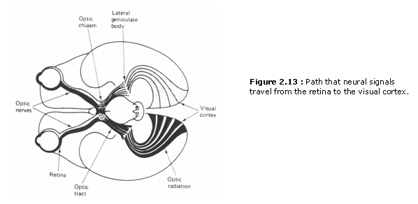
After the optic nerve bundles leave the two eyes. the
two bundles meet at an intersection called the optic chiasm. This is shown in
Figure 2.13. Each of the two bundles is divided into two branches. Two branches, one from each of the two bundles, join together to
form a new bundle. The remaining two branches form another bundle. This
crossing of the nerve fibers from two eyes is partly responsible for our
stereoscopic vision, which mixes the images from each eye to allow the visual
field to be perceived as a 3-D space. These two new bundles go to the left and
right lateral geniculate bodies, respectively. The
original fibers end here and new fibers continue to the visual cortex, where
the neural signals are processed and vision takes place. The visual cortex is a
small part of the cortex. a mass of gray matter
forming two hemispheres in the back of the brain. Little is known about how the
visual neural signals are processed in the visual cortex.